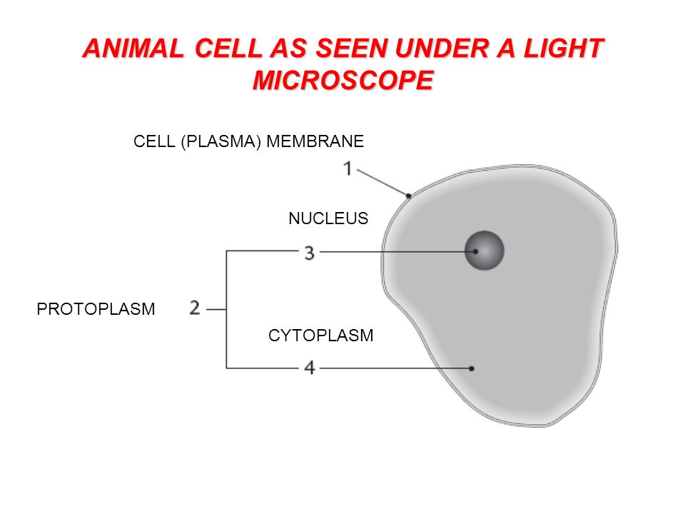
Animal Cell Seen Under Light Microscope : Centrosome Wikipedia / Under the light microscope, three parts can be seen in the animal cell:. Magnification, however, is not the most important issue in microscopy. This microscope introduces light that lets you view both living and dead cells through 2 lenses. A cell is a very tiny structure which exists in living bodies. Learn how to make an animal cell cake! Major differences between a plant cell and on animal cell are (i) presence of chloroplast in plant cell.
Be sure to include any structures that you notice in. 15 видео 74 483 просмотра обновлен 16 апр. (a)how is mitochondria adapted to its function? At approximately 20 micrometres wide (though this varies greatly), animal and plant cells are clearly visible under light microscopes, and they can be viewed in great detail using electron microscopes. What was once unseeable can now be seen, touched, and eaten!cut yourself a wedge for dessert or snack on a nucleus, lyosome, or…
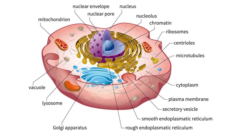
Here S How Plant And Animal Cells Are Different Howstuffworks from media.hswstatic.com Rana ray diagram of animal cell seen through electron. What was once unseeable can now be seen, touched, and eaten!cut yourself a wedge for dessert or snack on a nucleus, lyosome, or… (iii) presence of cell wall. The cell membrane, the structure that surrounds the cell and regulates the flow of substances between the cell and its surroundings; Given below is the diagram of a cell as seen under the microscope after having been placed in a solution Generalized structure of animal cell under light microscope. Line diagram of a general plant cell. .animal cell, as seen under a light microscope, limited to cell wall, nucleus, cytoplasm, chloroplasts, vacuoles and location of the cell membrane.
Present to a significant degree in animal cells) to generate contrast.
However, they usually can achieve a maximum of 2000x magnification which is not sufficient to see many other tiny organelles like ribosomes, endoplasmic reticulum, lysosomes, centrioles, golgi bodies unless they have an electron. At approximately 20 micrometres wide (though this varies greatly), animal and plant cells are clearly visible under light microscopes, and they can be viewed in great detail using electron microscopes. (ii) presence of large central vacuole in plant cell. Light microscopy (the use of microscopes is called microscopy), in plant cells c. The cell membrane, the structure that surrounds the cell and regulates the flow of substances between the cell and its surroundings; Central control, contains all information of chromosomes. Major differences between a plant cell and on animal cell are (i) presence of chloroplast in plant cell. The nucleus, usually spherical or ovoid structure that contains the genetic material; Terms in this set (8). The parts of a (palisade) plant cell that can be seen under a light microscope are:cell wallcell (surface) membranelarge (permanent) vacuolecytoplasmnucleuschloroplasts. There are one or more cells that form organism. Rana ray diagram of animal cell seen through electron. A cell is a very tiny structure which exists in living bodies.
I don't expect to see any cell wall, chloroplast. Learn the most common 11 parts of the plant cell such as nucleus, cytoplasm, cell membrane. Light microscopy (the use of microscopes is called microscopy), in plant cells c. Most cells are visible under a light microscope, but mitochondria and bacteria are barely visible. As for seeing electrons under any microscope in general, i would say we electron microscopes use accelerated electron beams (as opposed to visible light in a light microscope) to create images of magnification as high as 1 million x and has a very high resolving power to see the objects in fine detail.
Cell Structure Lesson Objectives By The End Of This Lesson You Should Know The Parts Of A Compound Light Microscope And Their Functions Pa Be Familiar Ppt Download from images.slideplayer.com Line diagram of a general plant cell. Central control, contains all information of chromosomes. What was once unseeable can now be seen, touched, and eaten!cut yourself a wedge for dessert or snack on a nucleus, lyosome, or… Image:plant cell seen under electron microscope. (iii) presence of cell wall. However, they usually can achieve a maximum of 2000x magnification which is not sufficient to see many other tiny organelles like ribosomes, endoplasmic reticulum, lysosomes, centrioles, golgi bodies unless they have an electron. You can see a variety of cells pretty well with the light microscope. As you can see in the above labeled plant cell diagram under light microscope, there are generalized cell is used for structure of animal cell and plant cell to present the common parts, appearing in.
This microscope introduces light that lets you view both living and dead cells through 2 lenses.
Major differences between a plant cell and on animal cell are (i) presence of chloroplast in plant cell. First seen with light microscopy 2. Transports substances such as protein. Under a microscope, plant cells from the same source will have a uniform size and shape. Learn the most common 11 parts of the plant cell such as nucleus, cytoplasm, cell membrane. (a)how is mitochondria adapted to its function? Now that we know what the main similarities are between all plant and animal cells, let's see how they when we view a specimen under a microscope it needs to let light pass through the specimen so we can see it. Animal cell cake of celliness: The nucleus, usually spherical or ovoid structure that contains the genetic material; Beneath a plant cell's cell wall is a cell membrane. Power house, provides cell with energy. Resolving power is the ability to distinguish between separate things which are close to each other. Be careful pushing it under the clips that the cover slide doesn't move or crack.
There are one or more cells that form organism. However, they usually can achieve a maximum of 2000x magnification which is not sufficient to see many other tiny organelles like ribosomes, endoplasmic reticulum, lysosomes, centrioles, golgi bodies unless they have an electron. Be careful pushing it under the clips that the cover slide doesn't move or crack. Raise the substage condenser to its top position and open the iris diaphragm all the there are three structures that distinguish plant cells from animal cells. What was once unseeable can now be seen, touched, and eaten!cut yourself a wedge for dessert or snack on a nucleus, lyosome, or…
3 from Learn the most common 11 parts of the plant cell such as nucleus, cytoplasm, cell membrane. First seen with light microscopy 2. Be careful pushing it under the clips that the cover slide doesn't move or crack. Beneath a plant cell's cell wall is a cell membrane. Rabies, seen here under a microscope, is an often fatal viral disease that a generalised animal cell as observed under an electron microscope. (ii) presence of large central vacuole in plant cell. The parts of a (palisade) plant cell that can be seen under a light microscope are:cell wallcell (surface) membranelarge (permanent) vacuolecytoplasmnucleuschloroplasts. Plant cells have cell walls, one large vacuole per cell, and chloroplasts, while animal cells will have a cell membrane only.
Be careful pushing it under the clips that the cover slide doesn't move or crack.
(i)mirror (ii)eye piece lens (iii)fine adjustment knob. We say cells are microscopic because they can only be seen under a microscope. (a)how is mitochondria adapted to its function? Plant cells have cell walls, one large vacuole per cell, and chloroplasts, while animal cells will have a cell membrane only. The cell membrane, the structure that surrounds the cell and regulates the flow of substances between the cell and its surroundings; Major differences between a plant cell and on animal cell are (i) presence of chloroplast in plant cell. Transports substances such as protein. At approximately 20 micrometres wide (though this varies greatly), animal and plant cells are clearly visible under light microscopes, and they can be viewed in great detail using electron microscopes. Most cells are visible under a light microscope, but mitochondria and bacteria are barely visible. Power house, provides cell with energy. Under a microscope, plant cells from the same source will have a uniform size and shape. The nucleus, usually spherical or ovoid structure that contains the genetic material; Line diagram of a general plant cell.
Share :
Post a Comment
for "Animal Cell Seen Under Light Microscope : Centrosome Wikipedia / Under the light microscope, three parts can be seen in the animal cell:"

Post a Comment for "Animal Cell Seen Under Light Microscope : Centrosome Wikipedia / Under the light microscope, three parts can be seen in the animal cell:"